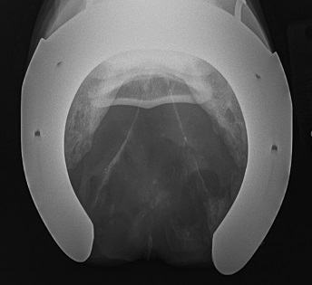
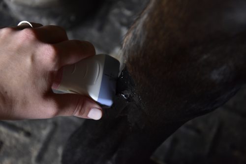
Excellent quality diagnostic imaging is what we are building our practice around. All of our equipment is portable and digital. We made our truck a mobile hotspot as well so we can send x-rays or ultrasounds anywhere in the world right from the barn aisle or show grounds, or to a specialist for review or an official report. All of our imaging is stored locally as well as backed up to the cloud, so we can always access it and know it is safe and secure.
Radiography
We carry the NEXT x-ray, a digital radiography system that allows us to acquire and view images instantly in the stall, barn aisle, or field. Our system allows us to radiograph the limbs up to the shoulder and stifle, back, neck and head in the field. We work with the Equine Medical Center of Ocala to image larger areas like the chest, abdomen, or pelvis which all require more powerful equipment. The EMC also performs bone scans and MRIs for our clients when needed. X-rays are used to evaluate bone, joints and teeth primarily. We can look for evidence of arthritis, trauma, fractures, infections and developmental orthopedic diseases, like OCDs.

Ultrasound
We utilize a GE Logiq E portable ultrasound to look at tendons, ligaments, and other soft tissues. Ultrasound uses sound waves to map soft tissues and show us a black and white image. We also utilize our ultrasound for performing guided therapies, where we can visualize placing a needle in a specific location at an injury or near a joint to provide medications directly to the injured area.

Prepurchase Radiographs
Our prepurchase radiograph set includes 38 radiographs of the front feet, front and hind fetlocks, hocks, and knees or stifles. Depending on the horse’s history and intended use, we will tailor the views to get the best information for you. If another veterinarian will also be reviewing the radiographs and has specific views they would like to see, let us know before the exam and we will include them. We are happy to perform more than the standard set of films, and can include other regions like the neck, back or shoulders at your request or depending on what we find during the exam. Our prepurchase films include a written report that details our findings, summarizes what the most important ones are, and discusses what the clinical impact may be of any changes seen on the radiographs. We pride ourselves on taking high quality images, so if your veterinarian or a radiologist is ever unhappy with the quality of a radiograph we submit, we will redo it at no charge.
Have a set of prepurchase films you would like another opinion on? We are happy to review and provide a written report on any sets of outside films you have. During lameness exams, we also can review older imaging, typically at no cost. Access to older radiographs often helps us know if radiographic findings are new or old and can help guide additional diagnostics and treatment.
Curo
 The CURO is a new diagnostic in veterinary medicine. It uses acoustic myography to give us scores of active muscles by detecting the vibrations produced during muscle contraction. We can evaluate muscular symmetry and fitness in real time while your horse is lunged or ridden. This is especially helpful for rehabilitation as we can visualize how your horse’s muscles are working during a therapy or exercise, so we can make adjustments to correct their movement and help prevent further injury. This is especially helpful for evaluating the hindlimb proximal suspensory ligament. We have a lot left to learn about this exciting modality and how it can help us improve veterinary care. Dr. Jill is actively involved with researching the CURO and finding differences between healthy and injured horses, and the effects our protective gear and therapeutic techniques have on muscle activity. For more information, visit https://curo-diagnostics.com.

